Scientific Photography
Scientific photography demands precision and objectivity: exact documentation for research, education or legal evidence.
Scientific photography documents for research, education or legal evidence, and that imposes three conditions on every shot: a reference scale in the frame, reproducible lighting and intact metadata. If one is missing, the image stops being data: it can't be measured or compared with the next one. Calibrated color and repeatability matter more here than any aesthetic decision.
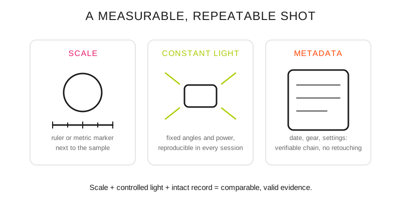
The three pillars of the scientific image: reference scale, reproducible lighting and intact metadata.
When does a photo become data?
In microscopy and laboratory photography the image is used to study samples with precision, ensuring that every capture is repeatable and comparable with others.
Archaeological documentation uses photography to preserve the state of pieces or sites before any intervention. Here the photo is a historical record that cannot be altered without a log.
In forensic and criminal photography the visual evidence must be objective and intact: every detail counts and any manipulation can invalidate its legal value.
Medical and clinical records require technical consistency so that a patient's changes over time are measurable.
In environmental and biological monitoring images make it possible to observe behavior patterns, cycles or ecological degradation.
In heritage conservation photography documents the state of works or buildings before, during and after restorations, with almost surgical precision.
What does each discipline require?
Discipline: Medical photography; Main requirement: Precise magnification; Critical condition: Exact color and uniform lighting
Discipline: Forensic documentation; Main requirement: Metric scale in the frame; Critical condition: Multiple angles, no interpretation
Discipline: Microscopy; Main requirement: Focus stacking; Critical condition: Specialized lighting, extreme magnifications
Discipline: Art conservation; Main requirement: Scientifically accurate color; Critical condition: Controlled, repeatable conditions
The stacking in microscopy is the same technique used in macro photography, applied at higher magnifications; and the exact color of medical records and artwork conservation starts from the principles of color theory.
What setup do I use for a scientific shot?
Calibration: calibrated professional monitor, specific color profiles.
Lighting: constant color temperature, calibrated to neutral white, no shadows. The setup of fixed sources and measured power is the same as in studio lighting, with reproducibility as the criterion instead of effect.
Scales: metric references in every shot, multiple magnifications.
Documentation: complete EXIF metadata, recorded conditions, strict protocol. Without orderly file management and workflow, the image's chain of custody breaks on the hard drive.
Common mistakes
Processing that alters relevant scientific information.
Inconsistent lighting between comparative samples.
Not including reference scales for measurement.
File compression that removes critical detail: the RAW is the original and the JPEG a working copy.
Professional tips
Scientific protocol: triple verification of calibration before each session.
Use specialized LED lighting with CRI above 95 for absolute color accuracy.
Document all technical conditions for future replication of the setup.
Frequently asked questions
What scale should I include in a documentation photo?
A rigid ruler or metric marker, placed on the same plane as the sample and parallel to the edge of the frame. If it's closer or farther than the object, the scale lies through perspective and later measurement comes out wrong; for pieces with volume it's worth repeating the shot with the marker at different heights.
Can a scientific image be edited?
Global, reversible adjustments (exposure, white balance, cropping) can be applied as long as they are recorded and the original file is kept intact. What invalidates the image is any local edit that adds, deletes or moves information: cloning, painting or recomposing. In the forensic field the rules of admissibility vary by country, so confirm the protocol of the jurisdiction before submitting.
Why is a CRI above 95 required in the lighting?
The color rendering index measures how closely colors under that light resemble those that would be seen under a reference source. Below 95, some tones (reds are usually the first) shift in ways that no white balance correction brings back, and two samples photographed with different lamps are no longer comparable.
What is needed for two sessions to be comparable?
Three things must be repeated: the same distance and focal length, the same position and power of the light sources, and the same color profile. Write down all three before dismantling the setup; without that record, the difference you see between the January session and the June one may come from the subject or the setup, and there's no way to know which.
Photography, when it is precise, becomes evidence.
The 44 Enfogram cards have the settings of each setup printed on them, so you can repeat it identically in the next session without depending on memory.